![]()
- LSE Center Publications
- Microscopy & imaging
- Super Resolution Microscope: Lattice SIM/STED/STORM
- Zeiss- Light Sheet Z7
- Nikon- Spinning Disk Confocal
- LSM 880- Upright confocal with MP laser
- LSM 710 – Inverted confocal
- LSM 700 – Inverted confocal
- LSM 980- Inverted confocal with Airyscan2
- Cytation™ 5 – BioTek
- Leica DMI8 Inverted Fluorescent Microscope
- Olympus Fluorescent Binocular Microscope
- Image analysis & processing
- Flow Cytometry
- Virus room – Emerson Building
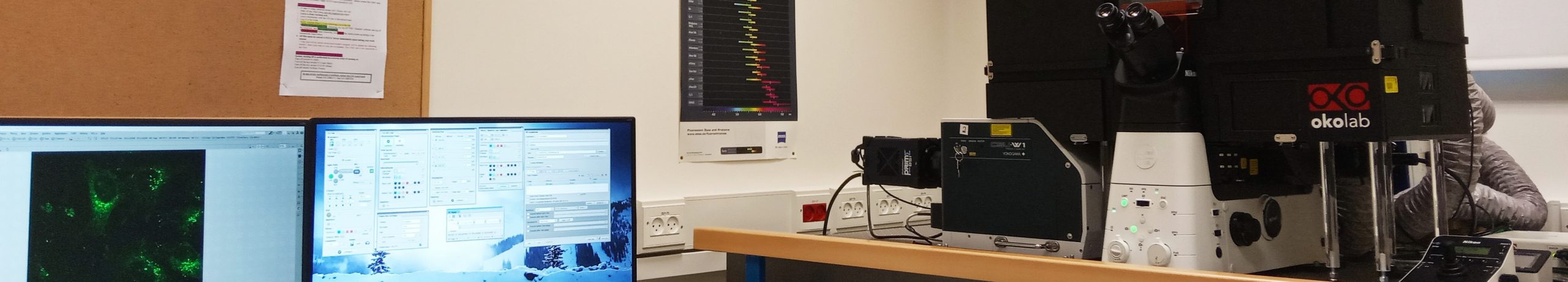
Nikon- Spinning Disk Confocal
Description
The Spinning Disk Confocal microscope from Nikon with CSU-W1 Confocal Scanner Unit with dual camera from Yokogawa, this system is designed for live-cell imaging.
It is hooked to an inverted fully motorized Ti2-E microscope stand.
Features
SYSTEM SPECIFICATIONS:
Imaging: Fast imaging with low phototoxicity;
Stand: Nikon Ti2-E
XY-stage: motorized
Z – Piezo focus NIDAQ 200 um range
Perfect focus
Sola LED illumination
Equipped for live imaging (temperature and CO2 incubation)
Software: NIS with image intelligence module
LASER OPTIONS:
The system includes 4 visible solid state lasers:
– UV diode laser – 405 nm (120mW)
– Blue diode laser 488 (200mW)
– Green diode laser – 561 nm (150mW)
– Red diode laser – 640 nm (200mW)
CAMERA SPECIFICATIONS:
Model: Two Photometrics BSI, sCMOS
Effective number of pixels: 2048 x 2048
Pixcel size: 6.5µm x 6.5µm
Effective Area: 13.312mm x 13.312mm
Dynamic range: 16 bit
Max frame rate: 43 fps @ 16-bit/ 12-bit (CMS)
63 fps @ 11-bit
Quantum efficiency (peak): > 95% (@560nm)
Read out noise: 1.0e- Read Noise with Correlated Multi Sampling (CMS) (depending on fps)
Spectrum Capabilities UV (200-400 nm), Visible (400-700 nm), Near Infrared (700-1000 nm)
OBJECTIVES:
| Magnitude | 10X – MRD00105 | 20X | 40X – MRD00405 | 60X – MRD07602 | 60X –TIRF – MRD01691 | 100X | 100X |
| Type | CFI PLAN APOCHROMAT LAMBDA |
CFI S PLAN FLUOR LWD |
CFI PLAN APOCHROMAT LAMBDA |
CFI PLAN APO VC 60XA WI |
CFI APOCHROMAT TIRF 60HX |
CFI SR HP PLAN APOCHROMAT LAMBDA S |
CFI PLAN APOCHROMAT |
| NA | 0.45 | 0.7 | 0.95 | 1.2 | 1.49 | 1.35 | 1.45 |
| Immersion | Dry | Dry | Dry | Water | Oil | Silicone | Oil |
| Working Distance | 4000 um | 2300 um | 130 um | 250 um | 130 um | 310 um | 230 um |
FILTERCUBES:
| LED FILTER | DAPI | FITC | TRITC | Cy5 |
| Ex | 392/23 | 474/27 | 554/23 | 635/18 |
| DM | 409 | 495 | 573 | 652 |
| EM | 447/60 | 525/45 | 609/54 | 680/42 |
CAMERA FILTERS :
| CAMERA | DAPI | FITC | TRITC | Cy5 |
| MASTER | 447/60 | 525/50 | 600/52 | 708/75 |
| SLAVE | — | — | 600/52 | 708/75 |
| Dichroic Mirror | for Dual camera | LP561 |
- Acquisition and analysis:
acquisition is performed using the NIS-Elements AR software which provides wide range of image processing functions: 2D/3D, projection, reconstructing, co localization, intensity measurements and more. The flexible secondary dichroic beam splitter makes this system fully capable of acquiring high resolution spectral scans and performing dual camera imaging. in addition, the NIS-Elements AR is optimized for advanced research applications. It features fully automated acquisition and device control through full six-dimensional image acquisition and analysis. The software is designed to deliver power and maximize workflow. It handles multi-dimensional imaging flawlessly, with support for capture, display, peripheral device control and data management and analysis of up to six dimensions (X, Y, Z, Lambda (wavelength), T, multi stage points). It also offers sophisticated image processing and visualization features, such as RAM capturing, HDR image capturing, intensity measurements over time, auto measurement, image filtering, macro and live comparison.
The system can also be operated in a “widefield” mode (Bypass the CSU) with superb sensitivity for faint fluorescence signal detection. Files generated by the Nikon-CSU can be loaded by the NIS Element viewer software which can by downloaded to every computer. Using the NIS one can process the images, create 3D rendering and export specific files to any other required form: JPEG, AVI, etc.
Applications
The system enables, amongst many others, the following applications:
* Fast Image Acquisition for Live Cell Imaging and Time-Lapse studies,
* Z-Stacks for localization of cells in living tissues and 3-D reconstruction
* High-resolution localization of sub-cellular compartments and quantities co-localization
* FRET – fluorescence resonance energy transfer
* Mark and find feature – The motorized stage allows marking specific coordinates using small magnifications or tiling of large scanning areas for further detailed scanning.