![]()
- LSE Center Publications
- Microscopy & imaging
- Super Resolution Microscope: Lattice SIM/STED/STORM
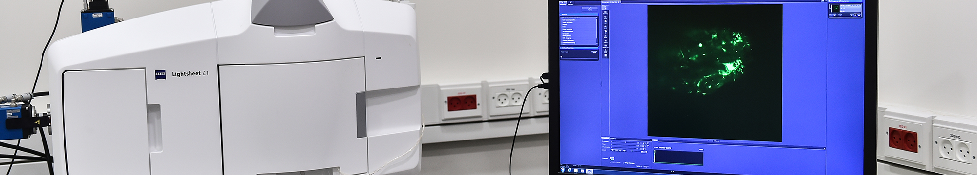
- Zeiss- Light Sheet Z7
- Nikon- Spinning Disk Confocal
- LSM 880- Upright confocal with MP laser
- LSM 710 – Inverted confocal
- LSM 700 – Inverted confocal
- LSM 980- Inverted confocal with Airyscan2
- Cytation™ 5 – BioTek
- Leica DMI8 Inverted Fluorescent Microscope
- Olympus Fluorescent Binocular Microscope
- Image analysis & processing
- Flow Cytometry
- Virus room – Emerson Building
Microscopy & imaging
The Microscopy core facility center contains: Multi-modality super resolutions microscope (SIM/STED/STORM), Light Sheet microscope, 4 confocal microscopes, High Content imaging systems (Cytation 5 & incell 2000), 2 inverted fluorescent light microscopes and 3 fluorescent binoculars. Additionally, there are work stations containing image analysis software, including Deconvolution software, time-lapse, 3-D reconstruction and other software.
All users receive full training on a specific instrument prior to independent work.
Workshops and hands-on sessions are organized to provide researchers with training in fluorescent and confocal microscopy. Additionally, a forum is managed for open discussions relating to new technologies, and sharing of ideas and personal experiences with the various systems.
The center contains the following microscopes:
High end microscopes:
- Elyra 7 eLS – Multi Modality Super Resolution (SR) microscope that combined lattice SIM, 2D-STED and STOTM/PALM methods for SR, this system is designed for live-cell imaging.
- Light Sheet Fluorescent Microscope– Zeiss Z7, with dual sided illumination, 2 cameras and full incubation. An analysis computer is available for the Light Sheet data analysis.
Confocal Microscopes:
- Spinning Disk Confocal microscope from Nikon with Yokogawa CSU-W1 Confocal Scanner Unit with dual sCMOS Prime BSI camera from photometrics, this system is designed for fast imaging and for long live-cell imaging.
- Confocal Zeiss LSM 980, an inverted confocal with 4 laser-lines, 2 PMT detectors, 32 ch GaAsp array detectors and Ariyscan2 (super-resolution) detector. Included full incubation, the system is designed for live cell imaging.
- Confocal Zeiss LSM 880, an upright microscope with 5 lasers, including a multi-photon laser and GaAsp and NDD detectors.
- Confocal Zeiss LSM 710, an inverted confocal with 6 laser-lines, 4 detectors and full incubation, dedicated for live cell imaging.
- Confocal Zeiss LSM 700, an inverted microscope with 4 lasers suitable for live cell imaging.
High Content Analysis system:
- Cytation™ 5 system combines automated digital microscopy, BioSpa incubator for up to 8 multi-dish plates and a robot machine.
Fluorescent Microscopes:
- Inverted Leica DMI8– inverted fluorescent microscope.
Fluorescent Binocular:
All users must download the User Statement and center policy and sign it before starting to work (See the Ordering section on this site, sub heading: User Statement and center Policy).
Microscopy Team:
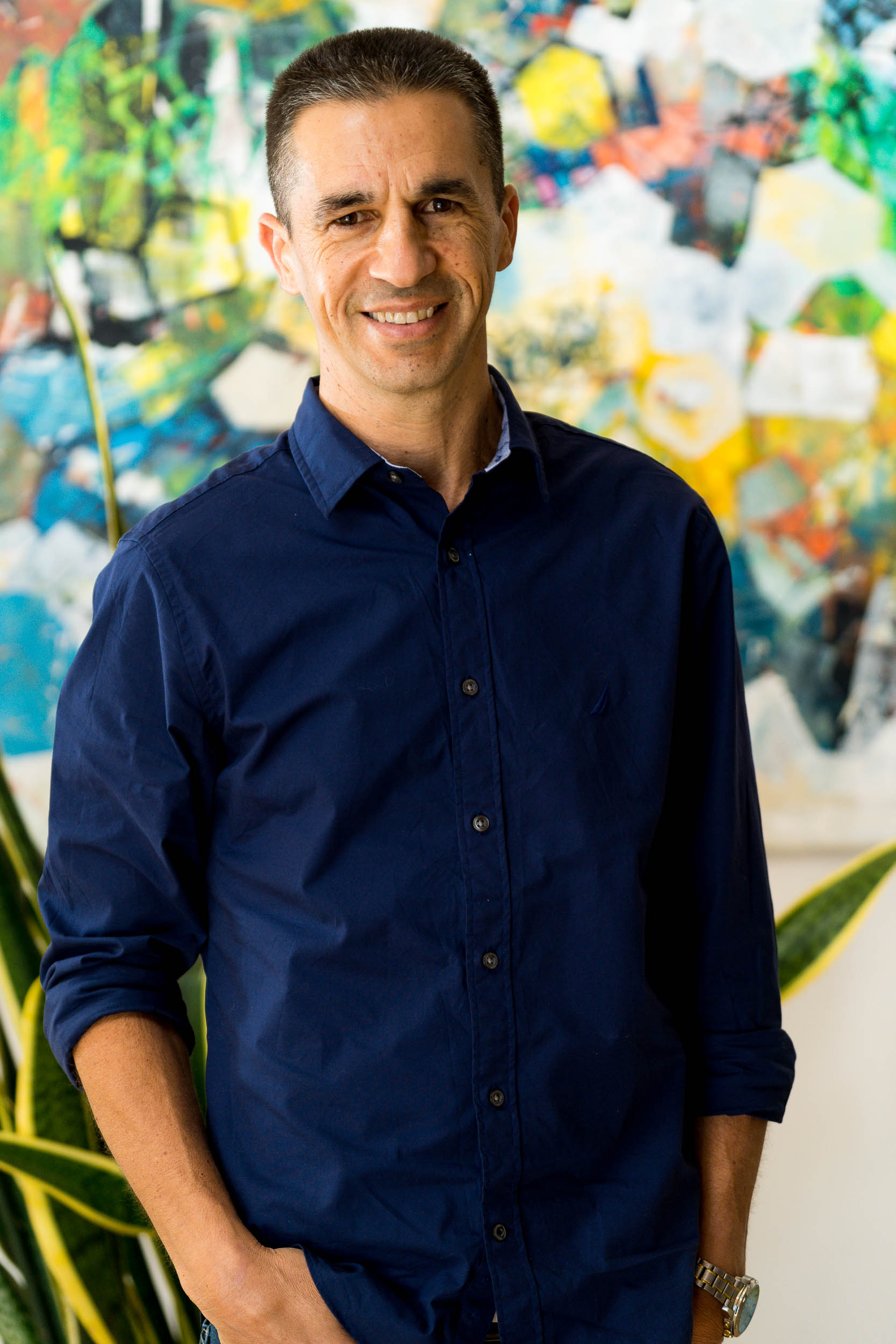
Dr. Nitsan Dahan
Microscopy & Analysis core facility, Head
Phone: +972 778 871 385 eMail: ndahan@technion.ac.il
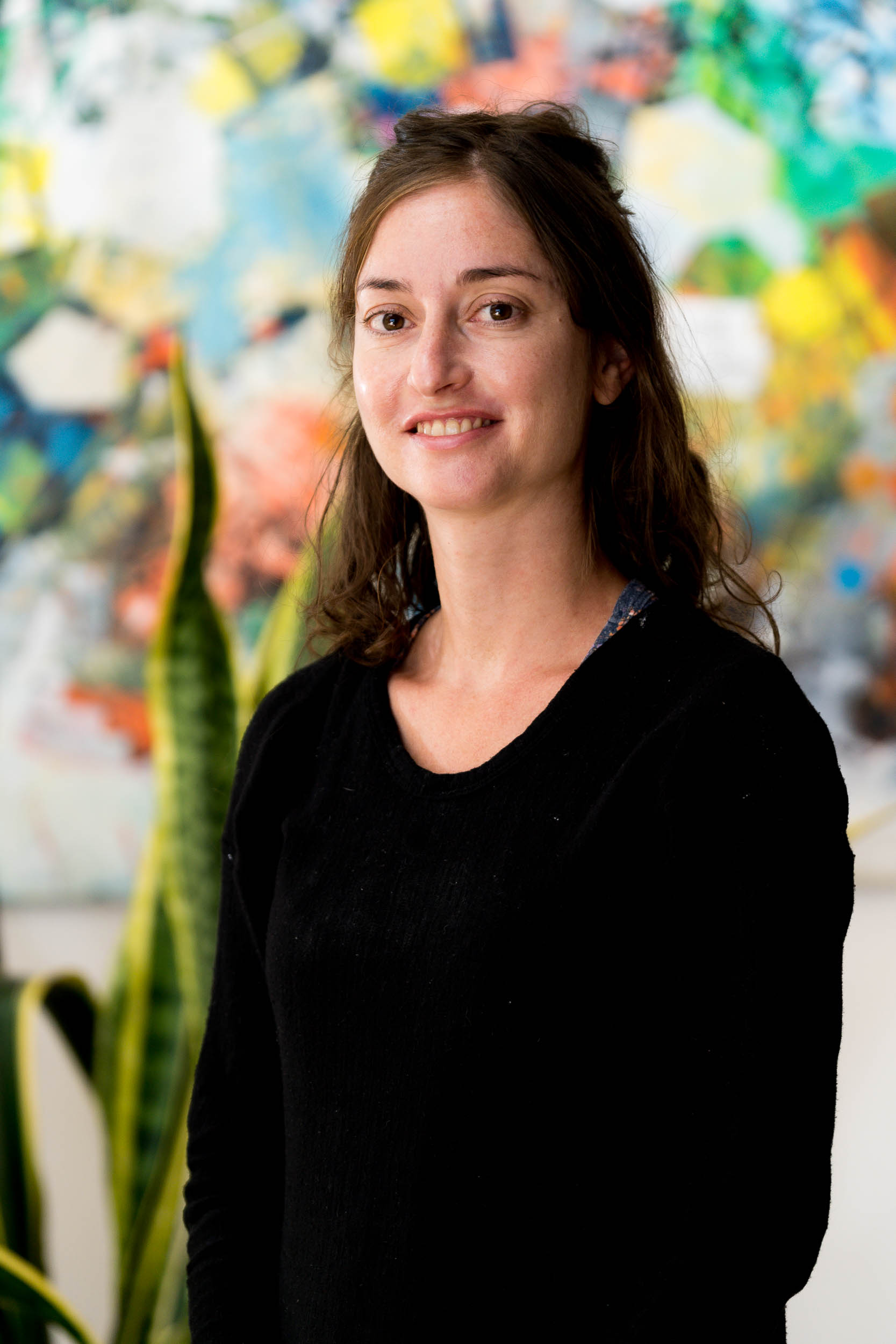
Dr. Yael Lupu-Haber
Bioanalyst & Microscopy Specialist
Phone: +972 77 887 1386 eMail: Yaelupu@technion.ac.il