![]()
- LSE Center Publications
- Microscopy & imaging
- Super Resolution Microscope: Lattice SIM/STED/STORM
- Zeiss- Light Sheet Z7
- Nikon- Spinning Disk Confocal
- LSM 880- Upright confocal with MP laser
- LSM 710 – Inverted confocal
- LSM 700 – Inverted confocal
- LSM 980- Inverted confocal with Airyscan2
- Cytation™ 5 – BioTek
- Leica DMI8 Inverted Fluorescent Microscope
- Olympus Fluorescent Binocular Microscope
- Image analysis & processing
- Flow Cytometry
- Virus room – Emerson Building
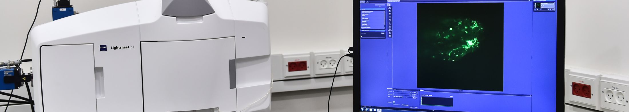
Zeiss- Light Sheet Z7
Description:
Light-sheet fluorescence microscopy (LSFM) is known also as selective/single plane illumination microscopy (SPIM). In this technique, the sample is illuminated by a thin sheet of excitation light (usually a few hundred nanometers to a few micrometers) which penetrates the specimen perpendicular to the axis of observation. Consequently, the entire plane of focus can be imaged simultaneously, generating an inherent optical section by exciting only fluorescence from the in-focus plane. Then, the emission light is collected by a sCMOS camera. This allows high speed optical sectioning imaging of large 3D samples (extremely faster than any other microscopy techniques) at subcellular resolution with high sensitivity and minimal photo-toxicity. Also the good sectioning capability reduces the background signal and thus creates images with higher contrast, comparable to confocal microscopy.
The sample inside the LSFM can be rotated and tilted, thus enabling multi-view imaging of the sample with improved axial resolution.
Dual-sided beam illumination with two aligned objectives complete the 3D imaging of the sample with high resolution.
The exceptional stability of Lightsheet 7 lets you observe living samples over extended periods of time – even days – with less phototoxicity than ever before. Light sheet 7 microscope is designed to image very large optically cleared specimens with subcellular resolution. The Lightsheet 7 have dedicated optics, sample chambers and sample holders to accurately adjust to the refractive index of your chosen clearing method, and then image your large samples, even whole mouse brains.
Features:
- Dual sided illumination
- 2 sCMOS cameras for acquisition of 2 channels simultaneously
- Full incubation with control over temperature and CO2
- Water Chamber
- Clearing chamber for using with 5x foc objective for cleared sample
- Detection objectives:
| Magnitude | 5X (Clearing) | 5X | 20X | 40X |
| Type |
EC Plan-APO 5×/0.16 Foc |
EC Plan –
NEOFLUAR 5x/0,16 |
W Plan-APO
20x/1,0 DIC |
W Plan – APO
40x/1,0 DIC |
| NA | 0.16 | 0.16 | 1.0 | 1.0 |
| Immersion | Dry | Dry | Water | Water |
Sheet lens:
| Magnitude | 5x (Clearing) | 5x | 10X |
| Type | illumination objective
10×/0.2 foc |
Illumation Optics
Lightsheet Z.7 5x/0.1 |
Illumation Optics
Lightsheet Z.7 10x/0.2 |
| NA | 0.2 | 0.1 | 0.2 |
| Immersion | Dry | Dry | Dry |
| Use with Detection
Objectives |
5x/0.16 foc | 5x | 20x, 40x |
Solid State Lasers:
| 1 | 2 | 3 | 4 | |
| Type | UV | Blue | Green | Red |
| Wavelength | 405nm | 488nm, | 561nm | 638nm |
| Power | 20 mW | 50 mW | 50 mW | 75 mW |
Epi-Fluorescence Filters:
| Filter Set # | 9310 | 9313 | 9316 | 9317 | 9322 |
| Fluorophore | DAPI – GFP | CFP – YFP | GFP – Cy3 | GFP – mCherry | Cy3 – DRAQ5 |
| Short Emission | 420-470 | 460-500 | 505-545 | 505-545 | 570-640 |
| Beam Splitter | LP 490 | LP 510 | LP 560 | LP 560 | LP 650 |
| Long Emission | 505-545 | 525-565 | 570-640 | LP 585 | LP 660 |
Applications:
Selected application fields of Lightsheet 7:
- Imaging a large (tile) optically cleared specimens with subcellular resolution.
- Morphogenesis and embryogenesis: Fluorescence imaging of spatial-temporal patterns within cells during embryogenesis of model organisms such as Zebrafish and Drosophila.
- Organogenesis and cell dynamics: Fast imaging of cellular dynamics, such as cell migration, cardiac development, blood flow, vascular development, neuro-development or calcium imaging.
- 3D cell cultures: Live imaging of 3D cell cultures, spheroids and cysts, tissue and organotypic cultures, 3D matrices of cells in polymers and scaffolds, 3D imaging of embryonic bodies and more. Images can be analyzed for cell migration, expression patterns and cell proliferation, cell localization, cell morphology and development of 3D cell cultures.
- Structural imaging of fluorescently labeled living specimens: Imaging detailed volume of fixed and live specimens, for example zebrafish, C-elegance, Arabidopsis root.
- Imaging of marine organisms: Fluorescence imaging of marine organisms such as ciona, squid, plankton and flatworms.
- Single cell imaging: Single cells primarily benefit from the low bleaching and can be studied for morphology, motility and biophysics of microtubule dynamics.
Analysis:
Additional computation system is available for the analysis of the Light Sheet data
Releted Links