- LSE Center Publications
- Microscopy & imaging
- Super Resolution Microscope: Lattice SIM/STED/STORM
- Zeiss- Light Sheet Z7
- Nikon- Spinning Disk Confocal
- LSM 880- Upright confocal with MP laser
- LSM 710 – Inverted confocal
- LSM 700 – Inverted confocal
- LSM 980- Inverted confocal with Airyscan2
- Cytation™ 5 – BioTek
- Leica DMI8 Inverted Fluorescent Microscope
- Olympus Fluorescent Binocular Microscope
- Image analysis & processing
- Flow Cytometry
- Virus room – Emerson Building
Amnis ImageStream®X Mark II
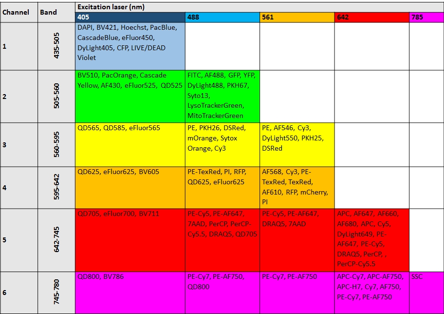
The Amnis ImageStream®X Mark II is an imaging flow cytometer equipped with 4 lasers. Imaging Flow Cytometry combines the speed, sensitivity, and phenotyping abilities of flow cytometry with the detailed imagery and functional insights of microscopy. The ImageStream® instrument produces multiple images of every cell directly in flow, including brightfield and darkfield (SSC), and up to 6 fluorescent markers.
Features
- Optics:
MultiMag – The MultiMag option for the ImageStream®X MKII system provides 60X and 20X objectives on a motorized stage, in addition to the standard 40X objective. The 60X objective offers greater resolution for the morphologic analysis of cells as small as yeast and bacteria, while the 20X objective offers a 120 micron wide field of view for very large cells. - EDF™ – The Extended Depth of Filed (EDF) option incorporates WavefrontCoding™ technology from CDM Optics, which is a combination of specialized optics and unique image processing algorithms, to project all structures within the cell into one crisp plane of focus. Ideal for automated FISH spot counting.
| Magnification | 20x | 40x | 60x |
|
Numeric Aperture |
0.75 | 0.75 | 0.9 |
| Pixel Size
|
1µm X 1 µm | 0.5 µm X 0.5 µm | 0.33 µm X 0.33 µm |
|
Field of View |
120 µm x 256 µm | 60 µm x 128 µm | 40 µm x 170 µm |
|
Imaging Rate |
4000 cells/sec | 2000 cells/sec | 1200 cells/sec |
Illumination:
Excitation – 405 nm, 488 nm, 561 nm, 642nm and Side scatter – 785 nm
High throughput autosampler:
-
- The autosampler option enhances productivity with unattended sample loading from 96 well plates. The workstation is managed by INSPIRE software.
Sample characteristics:
-
- • Volume: 20-200 μL
-
- • Utilization Efficiency: up to 95% of sample
-
- • Sample loaded in micro-centrifuge tubes
-
- Automated instrument operations:
-
- • Start up and shut down
-
- • Sample load and acquisition
-
- • Laser alignment, focus adjustment, calibration and self-test
Software
-
- INSPIRE® Acquisition Software – The INSPIRE® software for instrument setup, calibration, and spectral compensation. Intuitive user interface provides easy to use instrument controls, real time plotting and graphical gating, and images of every cell. Compensation wizard guides through multi-color compensation.
-
- IDEAS® Analysis Software – IDEAS® offers quantitative cellular image analysis and population statistics using standard or user defined features and masks. Numerical scoring of parameters such as size, shape, texture, co-localization and intensity allow quantification of visual data. Every dot on a scatter plot links directly to a cell’s images. Template and batch processing for automated data analysis for high throughput experiments.
Applications
Analysis of cells, yeast, bacteria, beads, and nano-particles. Fields of applications:
• Cell signaling
• Internalization
• Co-localization
• Microbiology
• Cell cycle
• Apoptosis
• Shape change
• Cell-cell interactions
• Oceanography
Releted Documents
INSPIRE® ImageStreamX MKII Software User’s Manual
IDEAS® ImageStream Analysis Software User’s Manual
ImageStream Applications Presentation


