![]()
- LSE Center Publications
- Microscopy & imaging
- Super Resolution Microscope: Lattice SIM/STED/STORM
- Zeiss- Light Sheet Z7
- Nikon- Spinning Disk Confocal
- LSM 880- Upright confocal with MP laser
- LSM 710 – Inverted confocal
- LSM 700 – Inverted confocal
- LSM 980- Inverted confocal with Airyscan2
- Cytation™ 5 – BioTek
- Leica DMI8 Inverted Fluorescent Microscope
- Olympus Fluorescent Binocular Microscope
- Image analysis & processing
- Flow Cytometry
- Virus room – Emerson Building
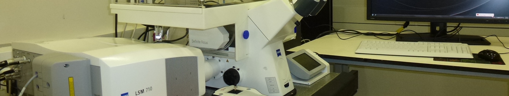
LSM 710 – Inverted confocal
Description
The LSM 710 laser scanning confocal microscope from Zeiss is a spectral imaging confocal system designed for live-cell imaging. It is hooked to an inverted fully motorized Axio Observer Z1 microscope.
Features
- Laser options:
The system includes 4 visible solid state lasers:
– UV laser – 405 nm (10mW)
– Argon multi line laser – 458, 488 and 514 nm (30mW)
– Green laser – 543 nm (10mW)
– Red laser – 639 nm (5mW)
-
Objectives:
| Magnitude | X10 | X20 | X40 | X63 |
| Type | EC Plan-Neofluar | Plan-Apochromat | Plan-Apochromat | Plan-Apochromat |
| NA | 0.3 | 0.8 | 1.2 | 1.4 |
| Immersion | dry | air | Water (correction ring) |
Oil DIC |
-
Controller: Two-mirror scanner system offers 360° XY scanning field rotation, quick zooming, different acquisition modes: simultaneous, sequential, bi-directional and more.
- Detection: The scanning module features a confocal beam path with 2 channels and continuously variable secondary dichroic beam splitter for free selection of emission bands; 2 symmetric galvanometer mirrors for high-precision beam positioning; 1 motorized and continuously variable high precision pinhole; 2 motorized emission filter wheels with 8 positions each, 7 of those can be exchanged by the user; 2 highly sensitive detectors (photomultipliers).In addition, the system provides transmission images via the Transmission Photomultiplier.
- Acquisition and analysis: acquisition is performed using the ZEN software which provides wide range of image processing functions: 2D/3D, projection, reconstructing, co localization, intensity measurements and more. The flexible secondary dichroic beam splitter makes this system fully capable of acquiring high resolution spectral scans and performing unmixing. The system can also be operated in a “filter-free” mode with superb sensitivity for faint fluorescence signal detection Files generated by the LSM 710 can be loaded by the ZEN LE software which can by downloaded to every computer. Using the ZEN LE one can process the images, create 3D rendering and export specific files to any other required form: JPEG, AVI, etc.
- Incubation: This microscope has an incubation chamber for live cell imaging for control over CO2, temperature and humidity.
Applications
The system enables, amongst many others, the following applications: Photoactivation and Photobleaching experiments with up to 99 freely definable ROI´s,
* Fast Image Acquisition for Live Cell Imaging and Time-Lapse studies,
* Z-Stacks for localization of cells in living tissues and 3-D reconstruction
* High-resolution localization of sub-cellular compartments and quantities co-localization
* Spectral imaging
* FRET – fluorescence resonance energy transfer
* Mark and find feature – The motorized stage allows marking specific coordinates using small magnifications or tiling of large scanning areas for further detailed scanning.
Related Links