Events

Save the Date # March 2ed 2023

The LS&E infrastructure center is happy to invite you to the LS&E Advanced Research Day. The day will take place at the Faculty of Biology Auditorium between 9:00 14:00 The day will include:  Students presentation Special guest lecture Advanced research applications at the LS&EMore details will be posted soon Hope to see you all, LS&E team ...

Continue Reading

LS&E Day for Image Processing & Analysis

On Dec. 25th @ Room 300 Biotechnology & Food Eng., Technion Agenda: 09:00-09:15 Gathering & coffee 09:15-09:30 LS&E infrastructure overview 09:30-11:00 Introduction to image processing & analysis 11:00-11:20 Coffee break 11:20-12:00 Advanced de-convolution (Dr. Douglas Lutz) 12:00-12:50 Image analysis tools (Fiji, Ilastik, Imaris, CellProfiler) & applications 12:50-13:00 Summary & New @ The LSE Registration is needed. Please register here LSE DAY for IA . For more information, please contact Dr. Yael Lupu Haber: yaelupu@technion.ac.il , 1386  ...

Continue Reading

April 25th, 10:30 – Seminars on Clearing methodologies for biological samples

The LS&E Microscopy core facility the invites You to attend special seminars in Advanced Microscopy Application and Methodologies. The next seminars will be on "Clearing methodologies for biological samples".   Date: Tuesday, April 25th, 10:30 o'clock, (45 min's talk) Location:  Room 4-17, Emersson Building Speaker: Shlomi Lazar, Ph.D. Division of medical chemistry, The Israel institute for Biological Research (IIBR). Title: Looking inside the organs: a comparative analysis of three methods for establishing optically transparent samples   Abstract: Typical histological study, using sectioning of the tissue, has major limitations in obtaining 3D images of structural components and cells distribution within tissues. Ideally, samples should be imaged at high spatial resolution with minimal sectioning. However, thick tissue imaging is limited mostly because of light scattering. In this study, we compared the efficacy of three recently published clearing protocols (Scale, 3DISCO and Clarity) in generating a transparent thick section which can be subjected to confocal analysis. Brains, ovaries and embryos obtained from transgenic mice expressing enhanced yellow fluorescent protein (eYFP) in...

Continue Reading
Microscopy seminar series

March 21st, 10:00 – Seminar on Insights into super-resolution

The LS&E Microscopy core facility the invites You to attend special seminars in Advanced Microscopy Application and Methodologies. The next seminar will be on "Insights into super-resolution – methodologies and applications of super-resolution microscopy ". Date: Tuesday, March 21st, 10:00 o'clock Please keep note that the seminar will be 30 minutes earlier than the original plan. Location: Auditorium , Faculty of Biology Speaker: Avi Jacob, Ph.D. Head of Light Microscopy facility, Bar-Ilan University.   Abstract: Microscopy-based methods for breaking diffraction limited resolution are collectively known as super-resolution (SR). There are several methods which can be divided into two main approaches: deterministic and stochastic. Stochastic methods include all localization techniques such as STORM and deterministic methods include the optical approaches such as STED. STED (STimulated Emission Depletion) is a mature technology, yet can require optimization to reach ~50nm resolution. Recently, companies have begun to offer "hardware-optimization + software based SR", whereby a measure of SR is achieved by automatically optimizing acquisition and then submitting to deconvolution....

Continue Reading

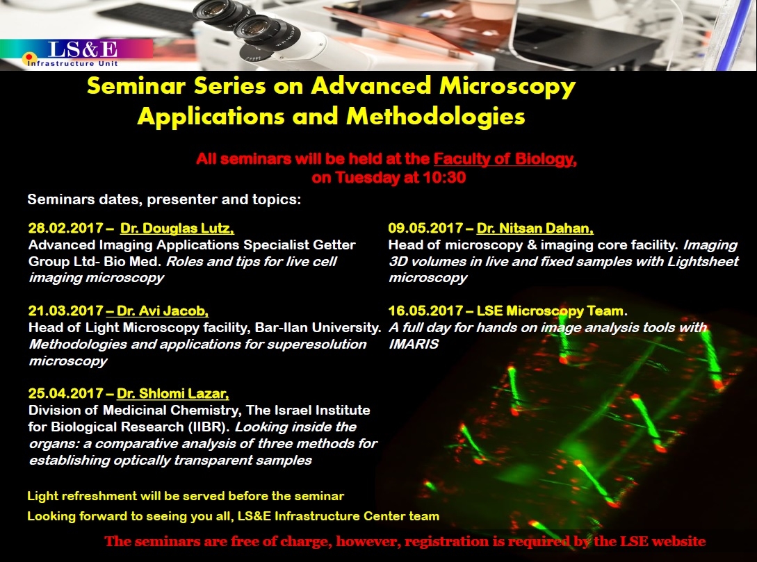
Seminar series on advanced microscopy applications and methodologies

  The LS&E Microscopy core is delighted to announce on seminars series on advanced microscopy applications and methodologies for Life Sciences Imaging taking place the biology faculty over the coming months. These seminars are designed as a program of short talks to enrich your knowledge on Live cell imaging, Clearing methodologies , Super-resolution and Image analysis aimed to help you optimize your microscopy imaging workflow. These seminars are free of charge, however, registration in event form  is required. Looking forward to seeing you all, LS&E microscopy core team.  ...

Continue Reading

Aquifer HIVE technology server platform is now available at the LS&E

The LS&E microscopy unit is happy to announce that HIVE technology is available for IMARIS and Light-sheet users. HIVE is a high speed centralized data repository that removes the need to constantly move or duplicate data sets in processing and analysis workflows The HIVE is a modular platform (computation & storage server) optimized for image processing and data handling. The HIVE designed for the pragmatic, biology-focused scientist, it boosts productivity in the LS&E microscopy unit .Its modular design integrates high speed processing, visualization, remote access, flexibility, data security, scalability and ease of use in one unit....

Continue Reading
Skip to content